Submission: Zhang Tianyu
Recently, the research team of A. Tittl, T. Lohmüller, and F. Keilmann from the Department of Physics at Ludwig Maximilian University of Munich (LMU), Germany, jointly developed a transient infrared nano-imaging technique based on scattering-type scanning near-field optical microscopy (s-SNOM), achieving label-free, non-destructive observation of the photoisomerization dynamics of individual lipid vesicles (as small as 176 nm) in aqueous environments, with a temporal resolution of 30 ms and a spatial resolution of 20 nm. For the first time at the single-vesicle level, the transient/delayed isomerization kinetics of azobenzene-based photoswitchable lipids (azo-PC) were revealed, and the correlation between their morphological changes (10% area increase, 8% circularity decrease) and chemical structure transformation was quantified.
This research was published in the internationally renowned academic journal Nature Communications under the title "Transient infrared nanoscopy resolves the millisecond photoswitching dynamics of single lipid vesicles in water". The corresponding authors are A. Tittl, T. Lohmüller, and F. Keilmann, and the first author is T. Gölz.
Research background
1. Importance of lipid nanocarriers: lipid vesicles (such as liposomes and lipid nanoparticles, LNPs) are core platforms of nanomedicine. They can encapsulate hydrophobic drugs for targeted delivery and have been applied in clinical scenarios such as mRNA vaccines (e.g., COVID-19 vaccines). Their performance optimization relies on externally triggered precise release mechanisms (such as light-controlled release), but this requires solving the challenge of observing dynamic processes with high spatiotemporal resolution.
2. Limitations of existing technologies: 1) Fluorescence labeling: azobenzene molecules quench fluorescence, interfering with the isomerization process. 2) Traditional microscopy: far-field infrared spectroscopy has low spatial resolution (~1 μm) and cannot distinguish individual vesicles; AFM, TEM, and the like can observe morphology but lack chemical specificity. 3) Dynamic monitoring gap: photoisomerization occurs on the millisecond scale, and existing technologies struggle to capture real-time structure-chemistry coupled changes in single vesicles.
3. Key challenge: a label-free, high-spatiotemporal-resolution, chemically specific technique needs to be developed to track the dynamic behavior of individual nanocarriers in real time in physiological environments.
Main innovations
1. Technical breakthrough — high-resolution chemical imaging of single vesicles in aqueous environments: 1) Spatial resolution: 20 nm (50 times better than traditional far-field infrared), with morphological details of 176 nm vesicles observed for the first time. 2) Temporal resolution: 30 ms (100 times faster than comparable near-field techniques), capturing millisecond-scale isomerization dynamics.
2. Methodological innovation — label-free dynamic monitoring: 1) No fluorescent or isotopic labeling is required; trans/cis states are directly distinguished through MIR fingerprint peaks (such as 1602 cm⁻¹), avoiding labeling interference. 2) Combining morphological (AFM topography) and chemical (infrared spectroscopy) data enables synchronous "structure-function" analysis.
3. Discovery innovation — nonlinear characteristics of isomerization kinetics: 1) Sudden acceleration phenomenon: trans→cis isomerization shows a rapid transition within 1 s after a delay of 10–15 s, possibly originating from cooperative effects of lipids within the membrane (disintegration of H-aggregates). 2) Morphology-chemistry coupling: cis-state vesicles show a 10% increase in area and an 8% decrease in circularity, directly related to reduced molecular packing density (25% volume increase of the cis form).
Research content and results
Fig. 1 systematically presents the experimental method and principle verification based on scattering-type scanning near-field optical microscopy (s-SNOM).
Fig. 1a is a working schematic of the membrane-based in-situ s-SNOM technique: a 10 nm thick SiN membrane separates the AFM probe from the lipid vesicles in the aqueous environment, which both avoids probe contamination and immobilizes the vesicles through van der Waals forces to maintain long-term stability. The MIR laser (red region) is focused at the probe tip to generate a localized near field, penetrating the SiN membrane to probe vesicles at a depth of 100 nm below; a UV/blue LED (blue region) uniformly illuminates the membrane surface to induce lipid photoisomerization. Fig. 1b supplements the experimental setup details, including the parabolic mirror focusing optical path and the Michelson interferometer design for detecting the scattered light amplitude (s2) and phase (φ2).
Fig. 1c presents the chemical composition of the vesicles: a 50:50 mixture of DOPC (a conventional lipid) and trans-azo-PC (a photoswitchable lipid), and the molecular structure schematic shows the conformational transition of azo-PC under 365 nm (trans→cis) and 465 nm (cis→trans) illumination — the benzene ring of the cis form changes from trans to cis configuration, with the molecular volume increasing by about 25%. The UV-VIS spectra in Fig. 1d verify the choice of switching wavelengths: the trans state has strong absorption at 365 nm, and the cis state has a characteristic peak at 465 nm.
Fig. 1e and Fig. 1f are the core experimental results. Fig. 1e is a spectrally averaged MIR amplitude image (s2) of a trans-state vesicle, clearly showing the outline of a vesicle about 500 nm in diameter (scale bar 500 nm). Fig. 1f compares the nano-FTIR phase spectra of the same vesicle in the trans (blue) and cis (purple) states, finding that the intensity of the benzene ring breathing peak at 1606 cm⁻¹ decreases significantly in the cis state — consistent with ATR-FTIR results — confirming the chemical specificity of this technique in distinguishing molecular isomeric states. In addition, the intensity of the carbonyl peak at 1742 cm⁻¹ shows no change, ruling out interference from laser power or alignment deviations.
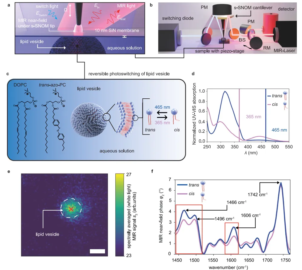
Fig. 2 focuses on the high-resolution imaging capability of s-SNOM for multiple vesicles, verifying the spatial resolution limit of the technique.
Fig. 2a and Fig. 2b show the MIR amplitude (s2) and phase (φ2) images of vesicles in a 15 μm × 15 μm region, respectively, at a probing wavelength of 1730 cm⁻¹ (carbonyl resonance). The results show that even in an aqueous environment, vesicles of different sizes (from 176 nm to several micrometers in diameter) can be clearly identified, and the phase signal (φ2 > 25°) is uniformly distributed inside the vesicles, with an approximately 100 nm wide fringe (white ring) at the edges.
Fig. 2c shows magnified phase images of four representative vesicles (scale bar 300 nm), and Fig. 2d extracts phase profiles along the vesicle centers and calculates the full width at half maximum (FWHM) through Gaussian fitting. The data show that the FWHM of the smallest vesicle is 176 nm, close to the system's spatial resolution limit (100–150 nm) and far superior to traditional far-field infrared microscopy (~5 μm).
This result confirms that s-SNOM can characterize the morphology of sub-200 nm nanocarriers in physiological environments. In addition, by comparing the experimental phase profiles with a theoretical model of a non-deformed sphere, it was found that the vesicles flatten on the SiN membrane — the membrane displaces upward by 1–2 nm (corroborated by mechanical images) — consistent with the near-field probing depth (~100 nm) being sensitive only to the 4 nm thick adsorbed lipid film, laying the foundation for quantifying vesicle morphological changes in subsequent dynamic studies.
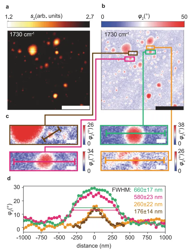
Fig. 3 visually demonstrates the dynamic morphological changes of vesicles induced by photoisomerization through time-series imaging.
Fig. 3a (amplitude s2) and Fig. 3b (phase φ2) record multiple switching cycles of the same vesicle at 1603 cm⁻¹ (azobenzene characteristic peak): the initial trans-state vesicle is circular; after 365 nm illumination (cis state), the morphology deforms significantly; and after 465 nm illumination it recovers (trans state). This process can be repeated for at least 4 cycles (within 1 hour), confirming the reversibility of the switching and the stability of the measurement.
Fig. 3c defines the morphological quantification metrics: area (A) and circularity (4πA/p², where p is the perimeter), where circularity = 1 indicates a perfect circle and smaller values indicate more irregular shapes. By segmenting the vesicle boundary with a threshold of s2 = 7.8 arb. units, Fig. 3d quantifies the morphological changes over two switching cycles: the trans→cis transition causes a 10% increase in vesicle area and an 8% decrease in circularity (from 0.85 to 0.78).
This result is consistent with the physical mechanism in which the increased volume of the azo-PC molecule in the cis state (20–25%) leads to reduced membrane packing density, and also supports the hypothesis that "the cis-state membrane has lower bending stiffness and deforms more easily". Notably, the morphological changes are synchronized with the spectral signal changes, indicating that s-SNOM can simultaneously capture the dynamic correlation between chemical structure (infrared spectra) and physical morphology (images), providing direct evidence for studying the structure-activity relationship of light-controlled nanocarriers.
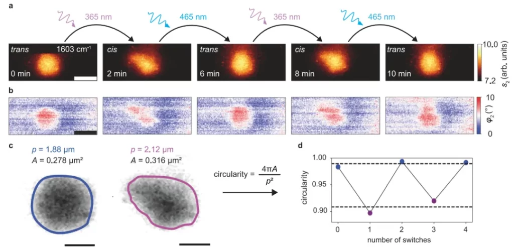
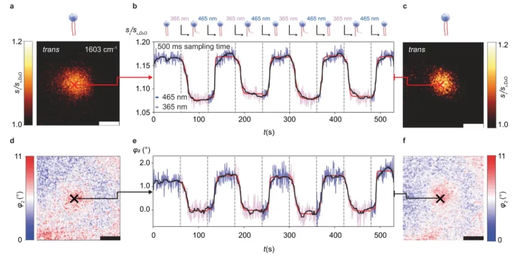
Fig. 4 reveals the millisecond-scale kinetic characteristics of photoisomerization through transient signal recording, breaking through the temporal resolution limitation of scanning imaging.
Fig. 4a and Fig. 4d show the amplitude/phase images (1603 cm⁻¹) of the vesicle before the experiment, with the probe fixed at the vesicle center (red cross) to avoid positional deviation. Fig. 4b and Fig. 4e show the signal time traces over 8 switching cycles (alternating 365 nm/465 nm illumination) with a sampling time of 500 ms: during trans→cis, the amplitude (s2) drops by 8% and the phase (φ2) decreases by 1.6°; during cis→trans, the signals recover symmetrically, with repeatability >95%.
Fitting the signal traces with a sigmoidal function (red curves) extracts key kinetic parameters: delay time td = 10.3–15.7 s (time from the start of illumination to the abrupt signal change) and growth parameter τ = 0.7–3.9 s (rate of the abrupt signal change). Since τ is on the same order as the sampling time (500 ms), the actual isomerization rate may be even faster. In addition, the abrupt signal change often occurs within 3–4 data points (1.5–2 s), suggesting a cooperative effect among lipids in the membrane — after the disintegration of trans-state H-aggregates, cis-state molecules rapidly rearrange, resulting in "delayed-burst" kinetic behavior.
Fig. 4c and Fig. 4f show imaging verification of the vesicle after the experiment, confirming no drift in its position or signal intensity and ruling out mechanical artifacts. Control experiments (D2O background, silicon wafer surface) further confirm that the signal changes originate from lipid isomerization rather than illumination interference. This method ultimately achieves a sampling time of 30 ms (signal-to-noise ratio 4σ), opening a new path for studying millisecond-scale biological dynamic processes.
Summary and outlook
This study provides a "visualization tool" for observing dynamic processes at the nanoscale. Its technical innovation (transient near-field infrared imaging in aqueous environments) and biological discovery (cooperative isomerization of lipid membranes) have cross-disciplinary inspirational significance for nanomedicine, biophysics, and materials science.
1. Core conclusions: 1) Technical validation: s-SNOM transient infrared nano-imaging enables label-free, high-spatiotemporal-resolution analysis of single vesicles in aqueous environments. 2) Mechanism elucidation: photoisomerization induces a transition of the lipid membrane from ordered (trans-H-aggregates) to disordered (cis-loose packing), leading to vesicle morphological remodeling and kinetic nonlinearity. 3) Quantitative data: spatial resolution: 20 nm; temporal resolution: 30 ms; isomerization delay time: 10.3–15.7 s; response rate: 0.7–3.9 s; vesicle area change: +10% (trans→cis); circularity change: -8% (trans→cis).
2. Limitations: 1) Membrane support influence: the SiN membrane may cause vesicle flattening (1–2 nm membrane displacement observed in experiments), requiring further optimization of the sample system. 2) Temporal resolution bottleneck: 30 ms is still limited by the detector response speed; in the future, brighter MIR light sources (such as synchrotron radiation) could push this to the microsecond level.
3. Future outlook: 1) Biomedical applications: extension to release kinetics studies of mRNA-LNPs, optimizing light-controlled drug delivery systems. 2) Technical extension: combining microfluidic chips to achieve real-time monitoring under dynamic environments (pH, temperature changes). 3) Cross-disciplinary value: providing a universal platform for dynamic studies of nanosystems such as metal-organic frameworks (MOFs) and 2D materials.
Paper information: Gölz, T., Baù, E., Zhang, J. et al. Transient infrared nanoscopy resolves the millisecond photoswitching dynamics of single lipid vesicles in water. Nat Commun 16, 6033 (2025). https://doi.org/10.1038/s41467-025-61341-9