
Author: Xu Xingxing
Background
In condensed matter, microscopic electric fields control most fundamental excitation processes and drive applications approaching terahertz (THz) frequencies in electronics. However, only a few imaging schemes can resolve subwavelength fields in the terahertz band, such as scanning probe techniques, electro-optic sampling, and ultrafast electron microscopy. Nevertheless, intrinsic constraints on sample geometry, acquisition speed, and field strength limit their applicability. This makes efficient and precise imaging of terahertz electric fields — especially field distributions at the nanoscale — an important scientific challenge.
To this end, Professor Georg Herink of the University of Bayreuth, Germany, and Professor Paul Mulvaney of the University of Melbourne, Australia, jointly published an article titled "Ultrafast imaging of terahertz electric waveforms using quantum dots". They developed a novel terahertz near-field microscopy imaging system, named Quantum-Probe Field Microscopy (QFIM). The system exploits the quantum-confined Stark effect (QCSE) to encode ultrafast electric near-fields into the luminescence of colloidal quantum dots (CdSe-CdS), combining far-field imaging of visible light with time-domain sampling of electric waveforms. This achieves compatibility between strong-field excitation and sub-micron resolution, providing a direct route to ultrafast field imaging of complex nanodevices.
Main text
QFIM is mainly based on the fluorescence excitation of quantum dots and the modulation of quantum dot absorption by an external electric field to detect the microscopic terahertz field strength distribution. As shown in Fig. 1 and Supplementary Figs. 1 and 2, a femtosecond laser (green light, pulse width 150 fs) illuminates the quantum dots to excite fluorescence at 645 nm, and a visible-light camera captures the fluorescence image of the entire bowtie antenna. When terahertz radiation arrives, its field strength changes the amount of green light absorbed by the quantum dots; subtracting the fluorescence image before terahertz excitation from the one after excitation yields the actual terahertz-induced result. Since the terahertz and green-light excitation sources originate from the same 1030 nm femtosecond laser, coherent detection can be achieved by varying the relative time delay.
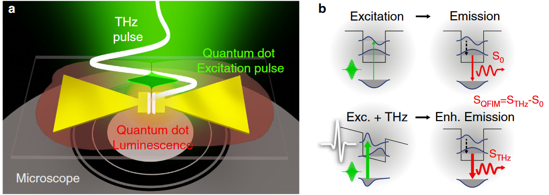
Fig. 1 Schematic of the QFIM principle. (a) A femtosecond laser (green light) and a terahertz signal simultaneously illuminate a bowtie antenna fabricated on a substrate bearing quantum dot material; (b) THz-induced changes in the quantum dot band structure increase absorption, which is converted into enhanced fluorescence emission.
The researchers fabricated a bowtie antenna above the quantum dots and illuminated it with both the femtosecond laser and terahertz waves. By varying the excitation delay between the two, they obtained microscopic snapshots of fluorescence emission at different delay positions, as shown in Fig. 2(b) — capturing not only the time-domain signal of the terahertz field, but also the ultrafast evolution of terahertz intensity over time. Although the luminescence lifetime of the quantum dots is very long (~10 ns), the time sampling resolution of QFIM is determined by the ultrafast absorption process, as can be seen from the transient absorption characteristics in Fig. 2(e).
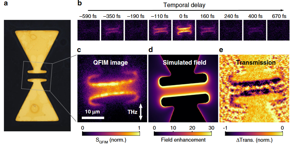
Fig. 2 Time-domain evolution of the terahertz resonant near-field in a bowtie antenna. (a) Optical micrograph of the bowtie antenna; (b) a series of microscopic fluorescence emission snapshots at specific delays, with the background without terahertz illumination subtracted; (c) transient fluorescence emission snapshot at 0 fs delay; (d) simulation result corresponding to panel c; (e) microscopic snapshot of the transient transmission of the femtosecond laser (green light) at 0 fs delay, showing the absorption characteristics of the quantum dots for the femtosecond laser, which is complementary to QFIM imaging.
By varying the peak value of the incident terahertz signal, the researchers also obtained the relationship between the terahertz field strength and the CCD-measured signal (Fig. 3(b)), thereby reconstructing the near-field waveform of the terahertz field at the bowtie antenna gap (Fig. 3(c)). Compared with the far-field terahertz signal obtained by electro-optic sampling, the spectral width is slightly reduced (Fig. 3(d)).
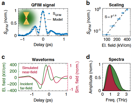
Fig. 3 QFIM signal and near-field waveform in the bowtie antenna. (a) Localized QFIM signal at the gap (blue dots) and the simulated fluorescence time-domain evolution based on the incident waveform (gray solid line); (b) QFIM signal peak as a function of the maximum incident field strength; (c) comparison of the driving far-field waveform (green) and the simulated local near-field inside the bowtie gap (red); (d) spectrum corresponding to the time-domain signal in panel c.
Finally, the researchers used this effect to observe terahertz plasmons excited by a 2 μm metal slit, as shown in Fig. 4(b), where the upper yellow region represents the direct uniform enhancement of the terahertz signal by the slit, while the lower white solid line represents terahertz plasmons excited by the slit with a phase velocity of about c/2. Fig. 4(d) shows the simulation results for these two delays: uniform enhancement at 0 ps and propagating terahertz generated at 1 ps.
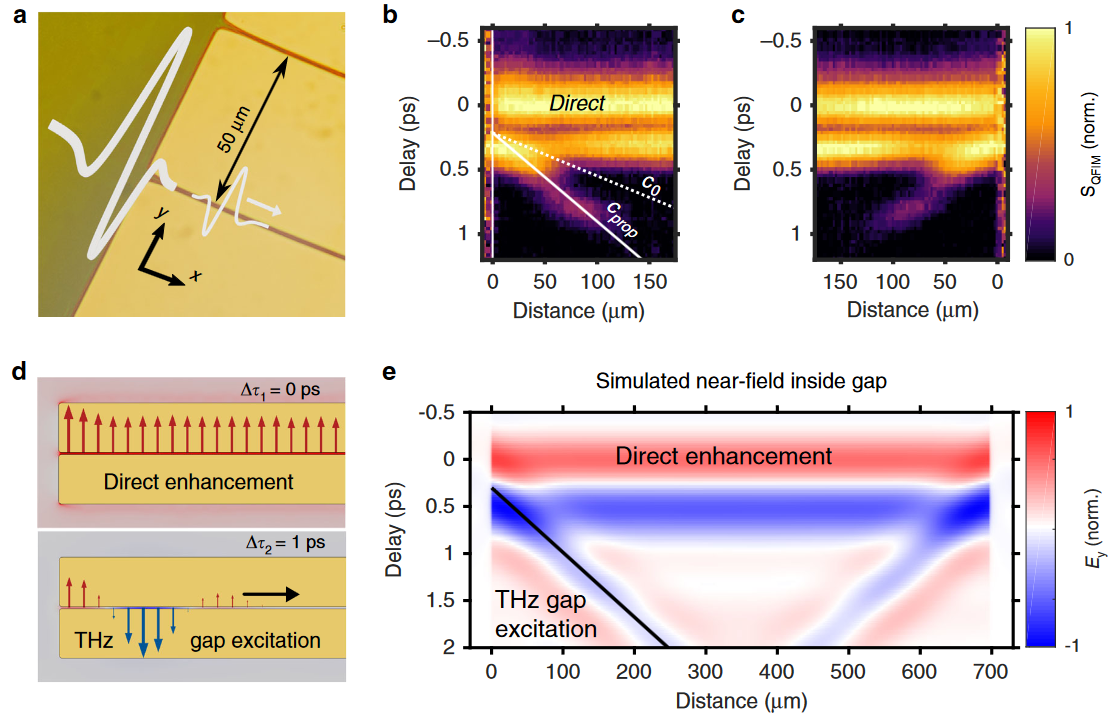
Fig. 4 Time-domain imaging of propagating terahertz gap excitation. (a) Terahertz waveguide excitation emitted in a microslit driven at normal incidence; (b) 2D display of the normalized QFIM waveform along the slit (position x, delay Δτ); (c) excitation on the opposite side of the waveguide, with the reversed propagation direction observed by QFIM; (d) simulated electric field distributions at two time delays illustrating the uniform direct enhancement (Δτ1 = 0 ps) and the excitation of propagating terahertz (Δτ2 = 1 ps); (e) 2D display of the simulated electric field evolution along the gap (x, Δτ), corresponding to panels b and c.
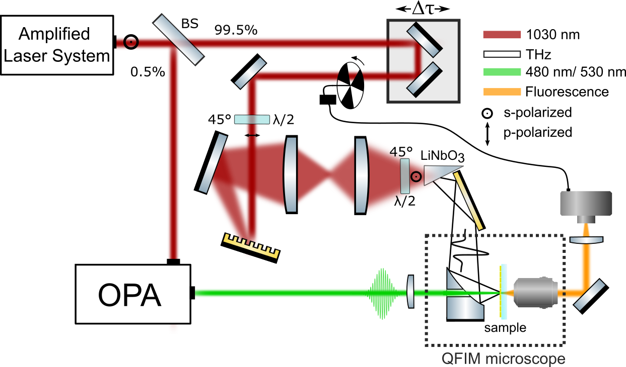
Supplementary Fig. 1 Schematic of the QFIM setup
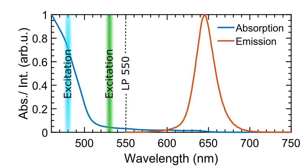
Supplementary Fig. 2 Absorption and emission characteristics of CdSe-CdS quantum dots
Summary
This method encodes instantaneous terahertz electric fields into the visible-light emission of nanocrystal quantum dots and far-field fluorescence imaging, thereby providing a direct link between luminescent observables and local electric fields for terahertz field driving and quasi-instantaneous QCSE. On this basis, time-resolved microscopic imaging of near-field waveforms inside a single bowtie antenna — a component of ultra-high-frequency devices, metamaterials, and strong-field light-matter interaction experiments — was achieved. Furthermore, the researchers observed terahertz propagation in a deep-subwavelength slit, introducing time-domain ultrafast sampling of electric fields propagating in confined structures. These results will trigger applications of QFIM in detecting electric field waveforms of surface excitations, including terahertz phonons and plasmons on bulk surfaces and two-dimensional heterostructures.
Note:
The quantum-confined Stark effect refers to the phenomenon in which, when an external electric field is applied perpendicular to a quantum well material, the absorption edge of the material shifts toward lower energies (red shift) as the field strength increases. Therefore, in conjunction with Supplementary Fig. 2, an increase in terahertz field strength leads to increased absorption of green light by the quantum dots.
References
Heindl, M.B., Kirkwood, N., Lauster, T. et al. Ultrafast imaging of terahertz electric waveforms using quantum dots. Light Sci Appl11, 5 (2022).