If you want to clearly see the optical response of a material at the nanoscale, scattering-type scanning near-field optical microscopy (s-SNOM) is a very powerful tool. It uses an ultra-sharp AFM probe to confine light to a region far smaller than the wavelength and then reads out local optical information near the probe, so many details that would otherwise be blurred into an indistinct patch under an ordinary optical microscope can be genuinely resolved.
But this brings its own challenge: many of the most interesting physical phenomena in quantum materials only emerge at low temperatures. To study these states, it is not enough to simply cool the sample—the entire near-field microscopy system must be brought into a cryogenic environment. It sounds like “adding a low-temperature chamber to a microscope,” but in practice it is far more complicated than that.
In 2022, Dapolito et al. published an intriguing work in Applied Physics Letters: instead of continuing to add more complex modules to cryogenic s-SNOM, they reversed the question—could any part be omitted directly? The answer lay in the AFM probe. They employed a self-sensing Akiyama piezoelectric probe, allowing the probe to directly “report” its own vibration state, thereby eliminating the optical system traditionally used to detect cantilever vibration in AFM. On this basis, they achieved cryogenic near-field imaging down to about 15 K.
The highlight of this work is not adding an entirely new imaging principle to s-SNOM. Quite the opposite—it does something seemingly unassuming: subtracting from the instrument.
In cryogenic s-SNOM, the first challenge encountered is “space anxiety.”
An s-SNOM system is by no means a simple microscope. It requires stable AFM probe scanning on the sample surface, precise focusing of infrared light onto the probe tip, and collection of extremely weak near-field scattered signals. In a low-temperature environment, these components originally spread out on an optical table suddenly have to be squeezed into a very limited space: the sample must be movable, the probe must be able to approach, and light must get in and out smoothly. In addition, traditional AFM typically also requires a dedicated optical detection system to monitor in real time how the cantilever is vibrating.
Thus, the cryogenic chamber quickly becomes crowded. As mentioned in the paper, a traditional cryo-SNOM often needs to accommodate the sample scanning and positioning mechanism, the near-field optical system, and the probe vibration detection device at the same time; the probe, sample, and optical components may also each require independent adjustment, so the entire system naturally becomes increasingly bulky.
Space, however, is only the first hurdle. Low temperatures also amplify many issues that would otherwise be barely noticeable: optical windows introduce additional heat, more mechanical components mean a greater cooling burden, and the probe itself becomes harder to tune after cooling. AFM control that is already well established at room temperature often needs to be readapted in a cryogenic environment.
So the real challenge is not simply “lowering the temperature,” but how to make the optics, scanning, low-temperature system, and mechanical control all work properly at the same time within a limited space. In this situation, eliminating even a single component can be very valuable.
What if the probe could "speak" for itself?
How does a traditional AFM know whether the probe is vibrating properly? One common approach is to direct another laser beam onto the back of the cantilever and determine the probe’s vibration state from changes in the reflected light. This can be understood as: the probe does the work, while a separate system has to be arranged alongside specifically to “watch it.” In an ordinary laboratory environment, this is not a big deal; but once inside a cryogenic chamber, an extra beam, an extra detector, and additional adjustment structures mean more space, more complex alignment, and extra thermal load.
The Akiyama probe used by Dapolito et al. offers another approach. This probe combines a microfabricated cantilever with a quartz tuning fork. Quartz is inherently piezoelectric, so when the probe vibrates, it directly generates a corresponding electrical signal. In other words, there is no need to separately “watch” the cantilever to know how it is vibrating.
Thus, the vibration detection chains of the two approaches can be written intuitively as follows: the conventional approach is “probe vibration → laser detection → obtain vibration information → adjust AFM”; after switching to Akiyamaprobe, it becomes “probe vibration → directly read the piezoelectric signal → adjust AFM”.
Thus, the optical path originally responsible for detecting the cantilever's motion can be removed from the system. This is where "self-sensing" becomes truly interesting: the probe not only scans the sample surface, but it can also incidentally tell the system 'how I am vibrating right now.'
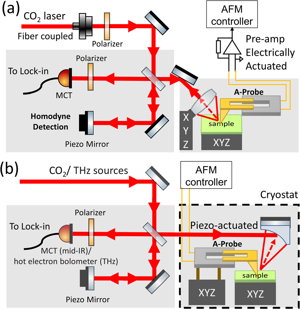
It might seem like just one less optical path, but for a cryogenic microscopy system, this change is far from trivial. Once the probe can perform vibration detection on its own, the layout of the entire instrument gains much more flexibility for rearrangement.
Less adjustment, more stability.
In the cryogenic setup presented in the paper, the authors further adopted a relatively compact optical layout. One typical choice is the use of fixed off-axis parabolic mirrors to focus the infrared light. From the perspective of conventional optical experiments, fixing the mirrors may not necessarily be a good thing: if the mirrors are adjustable, one can continue searching for a better focus position during experiments; once fixed, flexibility naturally decreases.
But what low-temperature instruments often pursue is not "everything can be adjusted." Fewer degrees of freedom sometimes actually make it easier to stabilize. Fixed optical components can make the structure more compact, and also easier to firmly connect with the low-temperature system; as temperature changes, component positions are less likely to drift, and the light spot is easier to keep stable.
This trade-off is quite illustrative of the design philosophy behind instruments for extreme environments. On a room-temperature optical platform, an additional movable component usually means one more way to optimize the system; but in a cryogenic system, it also means more space, a greater cooling burden, and another potential source of vibration and drift.
Therefore, evaluating a cryogenic microscope should not focus solely on how many adjustments it offers. Often, what really matters is determining which components must be retained and which degrees of freedom can actually be sacrificed.
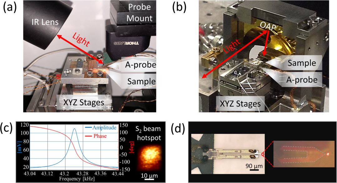
Image source: Dapolito et al., Applied Physics Letters 120, 013104 (2022), original paper Fig. 2.
From this perspective, the greatest value of “eliminating one optical path” is not just that it makes the setup look cleaner. What it truly saves is one of the most precious resources in low-temperature experiments: space. And this margin directly affects whether the instrument can continue to accommodate new experimental conditions in the future.
After all these subtractions, can it still be seen clearly at 15K?
Of course, no matter how compact the instrument is, it ultimately comes back to the most fundamental question: how good is the imaging? To verify this approach, the authors tested it on a standard SiO₂/Si sample. The results show that whether at room temperature or further cooled to 15 K using a cryogenic system, the Akiyamaprobe can perform both AFM topography scanning and near-field imaging, and the SiO₂ and Si regions remain clearly distinguishable in the near-field images.
This step is important because it demonstrates that the Akiyamaprobe does not simply make the setup “smaller”; it is genuinely capable of performing the scanning and near-field measurement tasks required for cryogenic s-SNOM.
However, low-temperature experiments did not become any easier as a result. As can be seen in the paper, after cooling to 15 K, stable acquisition of near-field signals is more difficult than at room temperature.
The reason is not mysterious: low temperatures alter the vibration characteristics of the probe itself, making the feedback control more sensitive. In other words, as the sample is cooled, the “mechanical system” inside the microscope changes as well.
This is also a reality often faced in low-temperature scanning probe experiments: lower temperatures do not mean the instrument becomes easier to operate—in many cases, the opposite is true. Experimenters must both investigate what low temperatures have changed in the sample and constantly verify what low temperatures have done to the probe and the instrument itself.
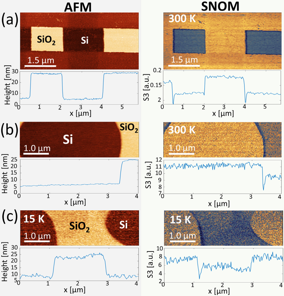
Image source: Dapolito et al., Applied Physics Letters 120, 013104 (2022), original paper Fig.4.
Making Room for Future Extreme Environments
It should be particularly noted that this work demonstrates cryogenic s-SNOM and does not directly achieve near-field imaging under strong magnetic field conditions. What it truly provides is an instrumental approach worth further extending to extreme environments.
Low temperature is only the first step. If in the future we also want to incorporate stronger magnetic fields, lower temperatures, or extend into the far-infrared and terahertz bands, the space inside the microscope will only become more constrained: the magnet takes up space, the cryogenic structure takes up space, and the optical path must not be blocked either.
At this stage, a device that can perform multiple functions simultaneously becomes particularly attractive. The Akiyama probe is exactly this type of solution: it serves as the probe that scans the sample, while also using its own piezoelectric response to provide vibration information. Tasks that originally required “a probe plus an optical detection system” are now further integrated into the probe itself.
The authors of the paper also explicitly point out that this more compact scheme has the potential to be further extended to far-infrared and strong magnetic field environments, and they identify lower temperatures and magnetic-field-dependent s-SNOM as future development directions.
From this perspective, what is most noteworthy about this work is not that any single parameter set a new record, but that it demonstrates a more fundamental instrument design approach:
Facing increasingly complex experimental conditions, it is not necessarily the only option to keep adding components to the system. Sometimes, a more effective approach is to let a single device take on more tasks, and then remove the modules that previously occupied space and consumed resources.
Summary
Bringing s-SNOM into a cryogenic environment has never been merely a matter of "cooling down." The real challenge lies in making nanoscale scanning, near-field optics, and the low-temperature system coexist stably within a single setup.
Dapolito et al. adopted an Akiyama self-sensing piezoelectric probe, providing a very intuitive solution to this problem. The probe can directly feed back its own vibration state through piezoelectric signals, so the part of the optical system in conventional AFM specifically used to detect cantilever motion can be omitted. Based on this more compact design, the authors further performed near-field imaging experiments down to about 15 K.
This may seem like just a small “subtraction” to the AFM optical path, but for future near-field experiments at lower temperatures, stronger magnetic fields, and longer wavelengths, having one fewer optical path is precisely what yields extra space and degrees of freedom.
Sometimes, an instrument truly moves into more extreme environments not because it has been packed with more things, but because it has finally learned what can be left out.
Source:
DapolitoM.,ChenX.,LiC.,etal.Scattering-typescanningnear-fieldopticalmicroscopywithAkiyamapiezo-probes.AppliedPhysicsLetters,2022,120:013104.DOI:10.1063/5.0074804
Note: All images in the main text are cropped from the original figures of the above paper, and the figure numbers have been renumbered according to the layout of this article.
Originally published in Chinese on the WeChat official account "见微而知著-觅几科技" (Miji Technology). English translation by Chengdu Miji Technology Co., Ltd. Source: WeChat article