Author: Yu Shuchao
Conventional optical microscopes are limited by the diffraction limit and cannot achieve nanoscale spatial resolution. The advent of scanning near-field optical microscopy (SNOM) broke through this bottleneck by using near-field coupling of evanescent waves to effectively overcome the diffraction limit. In recent years, SNOM has developed rapidly in surface science research, but its subsurface detection capability has yet to be systematically verified. In nanoscience and technology, a large number of key functional structures are often covered by electromagnetically transparent protective layers, so there is an urgent need for a non-destructive subsurface characterization technique with high spatial resolution and chemical identification capability. The rise of scattering-type scanning near-field optical microscopy (s-SNOM) provides a brand-new technical pathway to solve this problem.
In 2005, R. Hillenbrand et al. first demonstrated that s-SNOM has subsurface imaging capability. Their research showed that when the covering layer is a relatively optically transparent and low-interference material (such as a polymer or silicon dioxide), the near-field signal generated by the probe can penetrate the covering layer, enabling effective detection of deeply hidden structures. They carried out experimental verification in both the visible and infrared bands, successfully confirming this physical phenomenon.
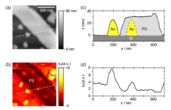
1. Visible light (633 nm wavelength): In the experiment, gold nano-island structures were fabricated on a silicon substrate surface and covered with a polystyrene (PS) film about 5 nm thick. The test results show that in the AFM topography image (Figure 1a), the PS film surface appears flat, while in the s-SNOM optical image (Figure 1c), the buried gold nano-island structures are clearly reproduced.
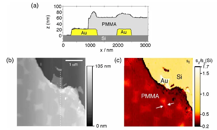
2. Infrared light (10.7 µm wavelength): When the gold nano-islands were buried beneath 50 nm of PMMA (Figure 2a), the experiment showed that the long-wavelength infrared light could not only clearly detect the subsurface gold islands 50 nm deep (Figure 2c), but also achieved a spatial resolution within 120 nm. This resolution corresponds to one-ninetieth of the working wavelength, far exceeding the imaging limit of conventional infrared microscopes.
This pioneering work was the first to prove that s-SNOM has non-destructive, nanoscale subsurface characterization capability. The technology can penetrate protective films, oxide layers, biological membranes and other media to directly image deep nanostructures, providing a brand-new means for studying buried interfaces.
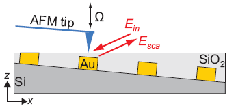
Although s-SNOM has subsurface imaging capability, the near-field signal decays sharply as the thickness of the covering layer increases. To address this, R. Hillenbrand et al. proposed two methods in 2012 to improve the signal-to-noise ratio and contrast of subsurface imaging. They used 100 nm-thick gold disks on a silicon substrate covered with a silicon dioxide layer of graded thickness as the research object (Figure 3), conducting experiments in the infrared band.
The first method is to reduce the tapping amplitude of the probe (Figure 4). Experiments showed that reducing the amplitude from 70 nm to 20 nm significantly improves the clarity and spatial resolution of subsurface imaging.
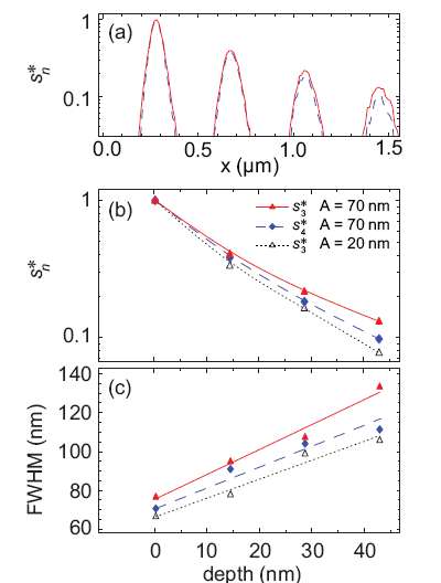
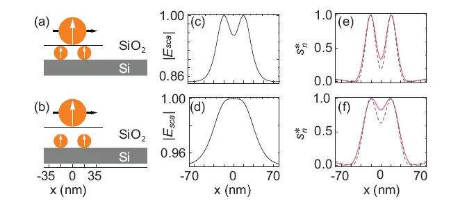
The second method is to use higher-harmonic demodulation. Higher-order near-field signals provide stronger subsurface contrast and can clearly resolve closely adjacent nanoparticles. At an amplitude A of 70 nm, the spatial resolution of the fourth-order signal is significantly higher than that of the third order (Figure 3b). The theoretical calculations in Figure 5 further confirm this phenomenon: Figures 5e and 5f compare the third-order (red solid line) and fourth-order (blue dashed line) near-field signal cross-sectional profiles of two adjacent nanospheres located directly below the surface and buried 15 nm deep, respectively.
In recent years, as the application potential of s-SNOM in materials analysis, biological imaging, semiconductor characterization and other fields continues to be realized, the demand from academia and industry for its subsurface imaging capability has become increasingly urgent. The theoretical and experimental methods proposed in the two pioneering papers above have extremely important guiding significance for subsequent subsurface near-field optics research.
References:
[1] Taubner T, Keilmann F, Hillenbrand R. Nanoscale-resolved subsurface imaging by scattering-type near-field optical microscopy[J]. 2005.
[2] Krutokhvostov R, Govyadinov A A, Stiegler J M, et al. Enhanced resolution in subsurface near-field optical microscopy[J]. Optics Express, 2012, 20(1): 593.